What makes PET/CT an essential diagnosis tool is the ability to provide accurate information about the body’s internal structures—by combining two different techniques. The examination is both safe and effective, which makes it a popular choice among patients.
What happens during a PET/CT?
As the name implies, a PET/CT scan uses two different techniques: positron emission tomography and computed tomography. That is why it belongs to the category of hybrid imaging techniques.
During the procedure, both the PET and the CT scanners are used to produce quality images, which are then combined to provide even more detailed information.
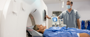
Regarding the order of examinations, the full-body CT scan is conducted first. Then the full-body PET scan follows. It is important that the patient lies still during both stages.
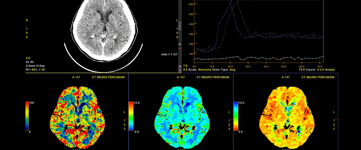
During CT, X-rays are used to create images of the inside of the body from different angles, which are then processed by a computer. This examination can provide information regarding the location, shape and size of a tumour. It can also detect the presence of other abnormalities.
During PET, a small amount of radioactive substance is injected into the patient’s vein. Depending on circumstances, the substance can also be taken orally or inhaled as a gas.
The ideal method of administration is chosen by the healthcare provider. After the substance is absorbed, it starts emitting gamma rays, which are detected by the PET scanner. The machines used to perform PET/CT scans are usually used for traditional PET scans as well.
How long does a PET/CT take?
A PET/CT can take anywhere between 30 minutes and an hour and a half to complete. This includes the preparation for both examinations, such as injecting radioactive substance into the patient’s vein.
Otherwise, the actual scan usually lasts between 20 and 30 minutes. If additional images are required, the scan can take longer.
Who is in charge of performing a PET/CT?
To efficiently perform a PET/CT, a team of healthcare professionals is needed. The most important member of the team is the radiologic technologist whose task is to operate the equipment and to make sure that the final images are of high quality.
A nuclear medicine physician or a nuclear medicine technologist must be present as well, as the examination requires the patient to get a radioactive substance injected. The images are then interpreted by a radiologist.
The collaboration of all these healthcare professionals ensures that the PET/CT is performed safely, correctly and efficiently—to provide the patient with the best care.
Preparations for PET/CT
Most of the details regarding a PET/CT procedure are discussed between the healthcare provider and the patient before the examination takes place. The patient must be made aware of both the benefits and the risks that come with this imaging technique.
One of the most common requirements is fasting for a certain amount of time before the scan. The fasting period is usually of about four to six hours. Otherwise, high blood sugar levels can interfere with the accuracy of the images, which makes it difficult to obtain a diagnosis.
It is also essential that the healthcare provider is aware of the medications or supplements the patient is taking, as some of them may affect the results of the examination.
As it is the case with any other imaging exam, relaxation is key: The patient should be advised to wear comfortable clothing and to remove any jewellery beforehand. More often than not, the patient is given a hospital gown to wear during the examination.
Advantages of PET/CT
- Complete results: Patients can obtain all the information they need, without having to undergo different examinations—as PET/CT combines both the PET and the CT scans. Completing the two examinations separately would take much more time.
- Accuracy: Thanks to the CT scanner, the exact location, size and shape of abnormal tissues can be detected during a PET/CT. This does not only lead to an accurate diagnosis, but to an efficient treatment plan as well. As both examinations are done at the same time, the patient does not have to change position, so there is less room for errors.
- Less radiation exposure: PET/CT uses a smaller dose of radiation than a simple PET, which makes it a safer examination for patients. As technology evolves, less radiation will be needed to obtain the information a person needs for a diagnosis.
- Versatility: The examination can be used to diagnose a wide variety of medical conditions—from detecting cancer, to determining the extent to which it has spread in the body, evaluating prognosis, assessing tissue metabolism, viability and many more.
Disadvantages of PET/CT
- Price: It is to be expected than a PET/CT is more expensive than other imaging techniques, as it is a combination of two different examinations. It is also worth to mention that insurance does not always cover the price of a PET/CT.
- Availability: Although MRI and CT scanners are common in imaging centres, the same thing cannot be said about PET scanners. The equipment is more expensive as well. Both the PET and the CT scanners are large machines, similar to a MRI unit. Despite that, some people might not be able to fit inside the machines because of their weight.
- Preparation: Undergoing a PET/CT requires a more thorough preparation than other imaging exams, including fasting before it.
To summarise, PET/CT comes with the advantages of both positron emission tomography and computed tomography, which makes it the ideal choice for patients who want to obtain as many information as they can about their health.


