Positron emission tomography or PET scans have revolutionised medical diagnosis and treatment in many ways by providing valuable information about the metabolic activity of the body’s tissues. For a more comprehensive information about the body’s internal structures, PET scans can be combined with other imaging techniques.
What happens during a PET scan?
During a PET procedure, the patient is supposed to swallow, inhale or be injected with a small amount of radioactive substance, also known as a tracer.
Then the tracer accumulates in the targeted area of the body, depending on the type of scan that is being done. Patients need to wait until the tracer is absorbed by the body, which can take up to one hour. The tracer emits radioactive signals, which are detected by the PET scanner.
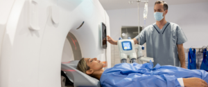
During the actual examination, multiple images are produced from different angles, which are then processed by a computer and transformed into detailed 3D images of the body.
This provides technologists with the insight they need about the cause of the symptoms the patient has been experiencing so far.
PET is mainly used to diagnose cancer, heart disease, coronary artery disease, Alzheimer’s Disease, seizures, epilepsy and Parkinson’s Disease.
How long does a PET scan take?
The actual scan can take 20 or 30 minutes to complete, depending on the area that is being examined. It is not unusual for an examination to take even longer, if the scan that is being performed is a difficult one.
If patients take into consideration the time it takes to prepare for a PET scan, then the examination can take anywhere between thirty minutes and two hours.
Who performs PET scans?
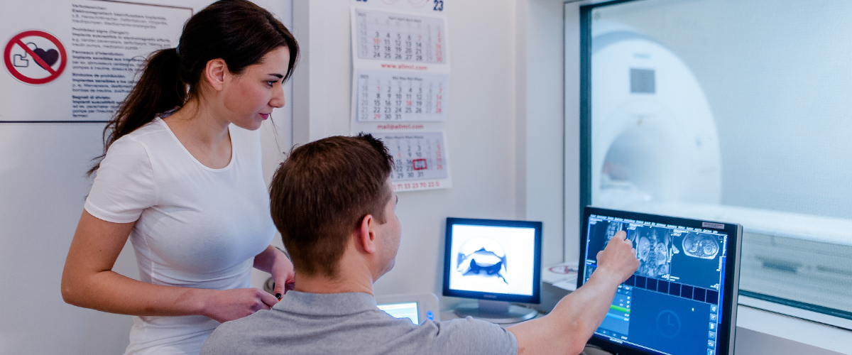
The scan is performed by nuclear medicine technologists who are professionally trained in the use of radioactive materials.
They don’t work on their own, instead they cooperate with other healthcare personnel, such as radiologists, so they can come up with an efficient treatment plan.
Which imaging techniques be combined?
Positron emission tomography can be combined with computerised tomography, and the result is a PET-CT. During this technique, the PET element is in charge of detecting metabolic activity, while CT provides useful information about the body’s anatomy.
This combination is especially useful when it comes to elaborating a personalised treatment. The PET scan can also be combined with magnetic resonance imaging, following a similar principle. These examinations are known as PET-MRI, and help monitor disorders.
Disorders can also be monitored through single-photon emission computed tomography (SPECT) scans, which use a different type of tracer than the traditional PET scans.
Advantages of PET scans
PET comes with a wide range of advantages that make them a valuable method in the diagnosis process, from being a non-invasive alternative to traditional examinations to accuracy.
- Non-invasive: Although injecting the tracer into the bloodstream is the most common way of performing a PET scan, it is not the only way. Depending on the patient’s situation, the agent can be given orally or inhaled as a gas. The ideal method of administration is determined by the radiology technologist who is handling the examination. But patients have the option to talk to them, so they know what to expect during the procedure.
- Early detection: Probably one of the most important advantages of PET is that it can detect changes in cellular activity in early stage, which allows patients to follow a treatment plan that can help them manage or even eliminate the symptoms they are experiencing in an efficient way. This is much more difficult to achieve, if the medical condition was left unchecked for a long time.
- Multiple applications: Although it is often used to identify cancerous tumours and determine the extent of the disease, PET can also be used to diagnose and monitor neurological disorders, heart diseases and infectious diseases. These examinations can also be used in the study of the effects drugs have on the body, which allows specialists to research new treatment options for patients.
- Accuracy: When it comes to tumours, PET scans provide useful information about their location and size, but also about the extent to which they have spread. After determining the stage of the disease, healthcare providers have all the information they need.
Disadvantages of PET
Just like any other imaging technique, PET has its own set of disadvantages:
- Radiation exposure: The dose of radiation that people are exposed to during the PET scan is small, but it is still there. In rare cases, it can increase the risk of cancer. Technologists can provide more information about the exact dose that is being used during the procedure, depending on the area that is being examined.
- Resolution: Subtle changes in tissue are less likely to be detected by PET when compared to CT or MRI scans which have much a much higher resolution. Small lesions are also difficult to see.
- Price: PET scans are significantly more expensive than other imaging techniques, which is why some people may hesitate when it comes to undergoing this examination. If PET is not covered by the health insurance, then patients have no choice but to pay the full price in exchange for the information they need.
- Limited availability: Not all imaging centres own the necessary equipment to perform PET. MRI and CT scanners are much more common than a PET imaging system, which is why it’s easier to choose one of those examinations.
To summarise, PET scans played a key role in revolutionising medical diagnosis and treatment. Without them, modern techniques such as PET-CT scans, PET-MRI scans or SPECT scans would not exist, which would make obtaining a diagnosis much more difficult for patients.
📷 Photo credits: daniela-mueller.com


